Workflows and Components of Bioimage Analysis: The NEUBIAS Concept

By Kota Miura, Perrine Paul-Gilloteaux, Sébastien Tosi, Julien Colombelli

DOI

NEUBIAS activities are spiraling around bioimage analysis “workflows”. Since this term is often used in slightly different ways by each person, we clarify the definition of “workflow” used in the NEUBIAS community in the following article.  While doing so we also introduce the related activities organized by NEUBIAS.


Software packages such as ImageJ, MATLAB, CellProfiler or ICY are often used to analyze bioimages. These software packages are “Collections” of image processing and analysis algorithms. Although their distribution and the way to access their resources is different since they can come with or without Graphical User Interface (GUI), as libraries such as ImgLib2, OpenCV, ITK, VTK, and Scikit-Image; we invariably refer to them as “Collections”. To actually analyze bioimage data scientifically and address an underlying biological problem, one needs to hand pick some algorithms from these collections, carefully adjust their functional parameters to the problem and assemble them in a meaningful order. Such a sequence of image processing algorithms with a specified parameter set is what we call a “Workflow”. The implementations of the algorithms that are used in the workflows are the “Components” constituting that workflow (or “workflow components”). From the point of view of the expert who needs to assemble a workflow, a collection is a package bundling many different components. Many plugins offered for ImageJ are mostly also collections (e.g. Trackmate, 3D Suite, MOSAIC…), as they bundle implementations of related algorithms.

Each workflow is uniquely associated with a specific biological research project because the question asked therein and the acquired image quality is often unique. This leads to a unique combination of components and parameter set. Some collections, especially those designed with GUI, offer workflow templates. These templates are pre-assembled sequences of image processing tasks to solve a typical bioimage analysis problem; all one needs to do is to adjust the parameters of each step. For example, in the case of Trackmate plugin for ImageJ, a GUI wizard guides the user to choose an algorithm for each step among several candidates and also to adjust their parameters to achieve a successful particle tracking workflow.  When these algorithms and parameters are set, the workflow is built. CellProfiler also has a helpful GUI that assists the user in building a workflow based on workflow templates, that allows the user to easily swap the algorithms for each step and test various parameter combinations.

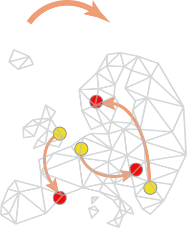
Though such templates are available for some typical tasks, collections generally do not provide helpful clues to construct a workflow – how and in which order the components should be assembled depends on expert knowledge, empirical knowledge or testing. Since the biological questions are so diverse, the workflow often needs to be original and might not match any available workflow templates. Building a workflow from scratch needs some solid knowledge about the components and the ways to combine them. It also needs an understanding of the biological problem itself. Each workflow is in essence associated with a specific biological question, and this question and the image acquisition setup affect the required precision of the analysis. In some cases, a higher precision does not imply more meaningful results, this should be carefully planned together with the statistical treatment (which also affect to some extent the choice of the components). Figure 1 summarizes the above explanations.


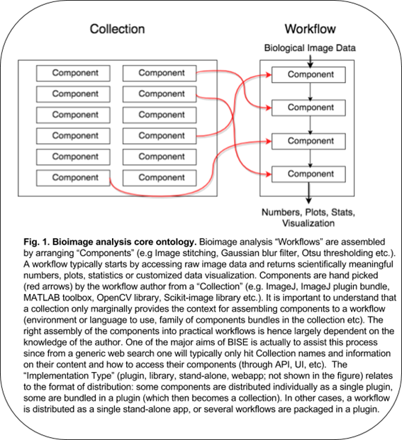
Many biologists feel difficulty in analyzing image data, because of the existing gap between a collection of components and a practical workflow. A collection bundles components without workflows, but it is often erroneously assumed that installing a collection is enough for solving bioimage analysis problem. The truth is that some expert has to choose components, adjust their parameters and build a workflow (Fig.1 red arrows), which is largely dependent on a priori knowledge. The correct assembly of components as an executable script is even more difficult in general, as it requires some programming skills. The use of components directly from library-type of collections, which hosts many useful components, also requires programming skills to access their API.  Bioimage analysts are then there to fill this gap but even they, who professionally analyze image data, need to always search for the best components to solve problems, reaching the required accuracy or coping with huge data in a practical time.

Another important aspect and difficulty is the reproducibility of workflows. We often want to know how other people are performing image analysis to learn new bioimage analysis strategies. We then try to find workflows addressing a similar biological problem. However, many articles do not document the workflows they used in sufficient details to reproduce the results. Some workflows are written as a detailed text description in Materials and Methods, but we recommend to publish them as executable scripts with documented parameter sets for clarity and reproducibility of analysis and results. As an extreme example, we found articles with their image analysis section in Materials and Methods merely documenting that ImageJ was used for the image analysis. Such a minimalism should be strictly avoided. For these reasons, we are promoting to publish bioimage analysis workflows in a reproducible format. The best format is a version-tracked script, i.e. a computer program because it is clear and reproducible. A script embedded in a Docker image is even better for avoiding problems associated with a difference in execution environments.

Many activities in NEUBIAS aim to address these difficulties, trying to make the process of choosing components and the construction of workflow easier, and to secure the reproducibility of published bioimage analysis workflows.

The workgroup 1 (WG1, Strategy and Scheduling) endorses the application of COST policies and moderates the strategic decisions taken in the organization of workshops and conferences. They promote communication among developers (who are implementing components and maintaining collections), bioimage analysts, microscopists and biologists, to increase the exchange of the usage information of the collections and workflows to bridge their gap (Figure 1 left and right).

The workgroup 2 (WG2, Training) aims at developing a multi-level training program in bioimage analysis based on the workflow-components concept. For beginners, basic components and their algorithms, as well as their useful assembly, are introduced. For intermediate level students, scripting languages are taught, to automate and to author reproducible workflows, and at the same time to learn practical workflows actually used in biological research projects.  For advanced level students, mainly comprised of professional bioimage analysts, advanced workflows and new components are introduced and studied in details for applications to a wider problem in biology and also to more efficiently author workflows.

The workgroup 7 (WG7, STSMs and Career Path) is organizing the extended and more individual aspect of such training, by supporting the travel of bioimage analysts and developers to other labs for implementing components and workflows in situ during missions of extended duration. WG7 is also making an effort to pave the career path of bioimage analysts, the novel type of profession in the life sciences community mediating computational science and life sciences.

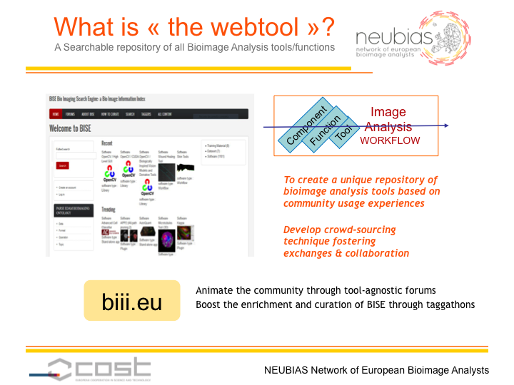
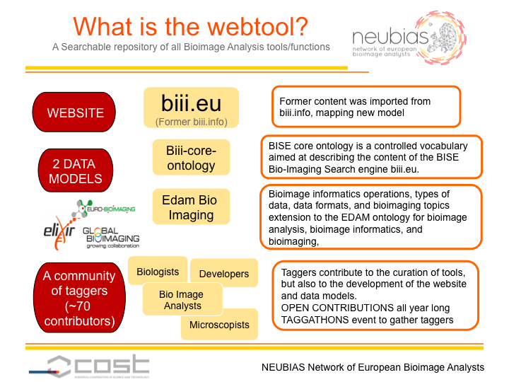
The workgroup 4 (WG4, The webtool BISE) is creating a searchable database of collections, workflows and components. General web search engines, such as Google, generally return hits of collections but not to the level of components. In addition, workflows are in many cases hidden in biological papers and difficult to be discovered. For these reasons, the webtool is expected to become a useful tool for those conducting the bioimage analysis.  The webtool is also designed to note impressions on the usability of components and workflows so that individual experiences can be swiftly shared within the community.

The workgroup 5 (WG5, benchmarking) is setting up a web tool enabling the interactive testing and benchmarking of some of the workflows from BISE. From this webtool the user will be able to select some specific annotated image dataset stored in an internal database and run compatible workflows on these images. The results from the analysis will be compared to the annotations (ground truth) to compute and display some problem specific performance metrics. The user will also be able to explore the results by interactively visualizing them overlaid on the original images. Since typically many workflows are available to solve a specific bioimage analysis problem, benchmarking them in such a unified environment is instrumental for fair comparison.

The workgroup 6 (WG6, open publication) aims at publishing the NEUBIAS teaching materials based on the workflow-components concept for a wider distribution outside of the NEUBIAS community, with detailed explanation on practical workflows associated with specific biological problems, for a better and more effective bioimage analysis in the biological community.

The workgroup 3 (WG3, outreach) works on communicating the outcome of all these activities towards wider scientific community, and also to promote communication among NEUBIAS working groups.

Overall, various activities of NEUBIAS are consistently directed: They all are reaching toward a clearer procedure for choosing components and for a more efficient, explicit and reproducible authoring of workflows for biological image analysis.


NEUBIAS social media, ‘What’s in it for me?’

by Auxiliadora Sarmiento and Irene Fondon (WG3)

There is surely no better place to meet and mingle with other professionals than at our community conferences. But in this increasingly connected world, more and more of our everyday personal interactions are taking place online. Thus, social networking sites are excellent tools not only for making but also for maintaining the connections between the members of the NEUBIAS community.

Nowadays, we have an active presence on the biggest three networking sites: Twitter, Facebook and LinkedIn. You might be surprised at the effectivity of these platforms for discussing science, forging collaborations and creating supporting networks. The thrilling thing about NEUBIAS social media is that you never know who may be able to help you.

 Twitter is a great way to expand your network well beyond the lab. An engaging conversation in less of 140 characters? Yes, of course. Our tweets communicate not just news but many other valuable contents, and the possibilities are huge. You can pose a question adding @NEUBIAS_COST and the NEUBIAS community will surely respond. If you can’t attend the last conference or symposium, don’t worry, you can search our hashtag #NEUBIAS and follow it in real time. And if you have a crazy result from an experiment, why not share it?

 Facebook is the biggest social network in the world. We mostly use it for sending out news, promoting upcoming events and publishing job openings. But you can also engage in interesting conversations about advances in imaging technology, or at least eavesdrop on them. You have an audience waiting, so share a link to your latest scientific manuscript or share a technique or a tool with the group.

LinkedIn is the Facebook of the professional world. People from different backgrounds are together: life scientists, bioimage analysts (BIAlysts), microscopists, developers and more. So, why not post an update of what you are doing? You can even become a thought leader by posting interesting content or promoting our own expertise.

Now you know our social media sites, it’s time to like us on Facebook, follow us on Twitter and join our network on LinkedIn in the case you had not done it before. Your contribution is valuable for NEUBIAS!! Do not be shy 😉

Inter-COST collaboration: NEUBIAS visited CHARME

By Natasa Sladoje (WG6)

COST’s mission is to support the integration of research communities. Inter-COST collaborations strongly contribute to that.
We have recognised an opportunity to establish such a collaboration with the COST Action 15110 CHARME – Harmonising standardisation strategies to increase efficiency and competitiveness of European life-sciences research (www.cost-charme.eu).
CHARME has 30 member countries, gathered to promote comprehensive, efficient, harmonized, recognized and adopted Standard Operating Procedures in Life Sciences. Observing that currently 80-90% of the research data are not reusable or accessible, CHARME aims at:
– identifying needs, developing norms and long-term strategy, and coordinating activities of the stakeholders (researchers, industry representatives and policy makers) towards standardization of acquisition, analysis, and publication of data;
– increasing confidence in quality of data to enhance sharing and re-use;
– supporting reproducibility of data and procedures.
CHARME states that “… standards assure and ensure that data become easily accessible, sharable and comparable along the value chain. The use of common standards may hence result in improved efficiency and competitiveness of European life-sciences research.”

NEUBIAS@CHARME’s Big Data in Life Sciences
NEUBIAS was presented during a 30min long invited talk at a training school (TS) and a workshop “Big Data for Life Sciences” co-organized by CHARME and EMBnet www.embnet.org in Uppsala (Sweden) September 18-22 (http://astrocyte.com/COST-CHARME/COST-CHARME/Home.html).
The audience was composed of the TS participants (students and trainers), invited guests
and the Action leaders. Host of the event was Erik Bongcam-Rudloff, the Action vice-chair. He, together with Domenica D´Elia, the leader of CHARME’s WG for dissemination and an EMBnet representative, showed high interest for NEUBIAS and possible further collaboration.

All activities of NEUBIAS attracted attention – our great success in training and education, our engaged communication, visibility and outreach via conferences, show-case events, STSMs, and other dissemination activities, and finally our several online tools and resources supporting and promoting data accessibility and integrity, availability of methods, reproducibility of results, and cooperation in a highly multidisciplinary domain. Several attendees expressed intention to join us in Szeged, at our 2nd NEUBIAS Symposium.

NEUBIAS is warmly invited to participate at the Workshop on Standardization in Life Sciences which will be organized by CHARME in Split (Croatia), October 23-25. http://www.cost-charme.eu/events/split

CHARME Training school on Big Data in Life Sciences attendees http://www.cost-charme.eu/news

10/13/2017

NEUBIAS Short-Term Scientific Missions

By Julia Fernandez Rodriguez and Clara Prats (WG7)

The Short-Term Scientific Missions (STSMs) grants are the best instrument NEUBIAS Action has to support ideas and knowledge exchange and collaboration among the network across borders. STSMs can vary from one week to three months. Both home and host institutions should be from a COST country which has accepted the NEUBIAS Memorandum of Understanding and has participated in the NEUBIAS Action.

Knowledge exchange – STSMs contribute to knowledge transfer to support individual careers and regional development. Alexander L. Hargreaves, from the University of Leeds UK, is one of many researchers who took part in a NEUBIAS STSM. His works involves a clinical trial investigating the effect of sperm DNA compaction faults on human fertility. Called HABSelect Clinical Trial, it’s the largest of its kind with over 3000 participants. Having only wide-field images in the Aniline Blue stain, Alexander performed a STSM in Uppsala, Sweden for one week to collaborate with image computation specialists from Prof. Carolina Wählby group. As champions of the popular open-source software CellProfiler, they have developed a range of image analysis algorithms, from shape-based thresholding and segmentation, to categorization with leading-edge deep learning architectures. “Thanks to the STSM, I appreciated learning a great deal in a short, high-pressure period of time from the very best scientists in their field. I also produced both valuable data and important future contacts for my home institution. Through NEUBIAS I feel we are forging important international links between BioImage Analysts and fertility experts across Europe. Overall, I hope that this large-scale study will act as a catalyst for improved image analysis throughout fertility science; eventually leading to better fertility outcomes for families”, says Alexander.

Cooperation and Collaboration – Enhancing further collaborations on innovative image analysis methods is also an important aspect of NEUBIAS STSMs. Victor E. A. Caldas from the Vrije Universiteit Amsterdam, performed an STSM at the laboratory of Prof. Mark Fricker, University of Oxford, UK. The specific goal of his STSM was to evaluate image datasets of symbiotic fungal networks, indicating directions for the next round of image acquisition and transfer technology of image analysis of living networks between their laboratories. “This STSM was fundamental to initiate the collaboration with Prof. Mark Fricker and to identify our common research interests and directions for future research. As a direct consequence of the STSM, Prof. Fricker will be included in my next grant application, aiming at optimizing image analysis of networks and modelling of adaptive fungal networks. I am convinced that this STSM has had a valuable and very positive effect in my research topic and career” says Victor.

NEUBIAS continues to see very positive results from these missions. Thanks to its flexible and bottom-up approach, researchers across Europe are working together to show impressive results in a short period of time!

How to Apply for a STSMs – BioImage Analysts and Life Scientists, from Research Labs AND Core facilities, can apply for funds to cover their expenses when visiting a Host-Lab in a different country (Lab or industry) where they will perform a short scientific project strictly focused on BioImage Analysis, and that should enable:
1) collaborations on innovative image analysis methods,
2) access to big data analysis technology and/or image analysis tools for scientists lacking them locally,
3) knowledge transfers to support careers and regional development.

Applications for the STSMs can be submitted any time and will be reviewed at the end of each month!

Please, check all the information here: https://eubias.org/NEUBIAS/mobility-grants/ and contact these people for any question:
Julia Fernandez Rodriguez – NEUBIAS STSM Coordinator juliafer@cci.sahlgrenska.gu.se
Clara Prats – NEUBIAS WG7 Co-Leader – cprats@sund.ku.dk
Julien Colombelli – NEUBIAS Action Chair – julien.colombelli@irbbarcelona.org

NEUBIAS taggathon – Building webtools for Bioimage analysis

By Perrine Paul-Gilloteaux, Giovanni Cardone, Florian Levet, Alban Gaignard, Julien Colombelli, Natasa Sladoje (WG4)
The general purpose of Taggathons, organized by WG4 in NEUBIAS, is to implement and feed the content of NEUBIAS webtool BISE (biii.eu).

This webtool aims to provide an organized repository of bioimage analysis workflows and software components to biologists, bioimage analysts and algorithm developers. Several goals have been identified for each user group:
For bioimage analysts: to offer a simple and integrated way to search and select components for constructing bioimage analysis workflows (such as scripts).
For life scientists: to provide a place where to easily find workflows that are close to their needs for completing image analysis tasks in their biological research projects.
For developers: to get up to speed on current available solutions, to discover alternative approaches to compare, and to identify gaps and needs of the community (find out what is missing in current bioimage analysis domain).
At the same time, all three user groups can actively contribute to the webtool by adding new pages, and by sharing with the community their own experience in using each of the component/workflow/collection.

To achieve these goals, the approach is to feed the content of the NEUBIAS webtool by crowdsourcing, based on a set of tools and guidelines developed during the taggathons. During the first NEUBIAS taggathon (Barcelona, 2016), a list of feature specifications has been ranked by importance, and potential web technologies have been reviewed and evaluated against these specifications. During the second taggathon, (Oeiras, 2017), new data models, both for the organization of the webtool content and the tags ontology, have been discussed in collaboration with NEUBIAS WG5, Eurobioimaging and Elixir, and have been implemented in the webtool. During the third taggathon (Gothenburg, 2017), content from the prototype (biii.info) has been reimported into the new webtool and has been curated in biii.eu. A new approach to improve the search experience of biologists has been discussed and is currently under implementation, through the introduction of relationships between terms in the ontology and common image processing wording used for defining biological problems (e.g filament tracing has neuron, microtubule, mitochondria, vessels as related terms). The next taggathon will take place in Hungary (Szeged, 2018) before the NEUBIAS Symposium, and will be focused on thematic curation of the BISE content focusing on Big-data generating techniques, e.g. Electron- and lightsheet- microscopy bioimage data analysis, but also on the core ontology updates and its use in BISE.

2nd NEUBIAS CONFERENCE

by Peter Horvath and Sebastian Munck (WG1)


After a successful first NEUBIAS conference in Lisbon last year, we are getting set for the next edition of the NEUBIAS conference, running from 27th – 02nd Feb in Szeged Hungary. We are excited to welcome you there.
Like last year the conference consists of two training schools, one for early career Investigators, and one for Bioimage Analysts. The Analyst course is organized by Kota Miura and Jean-Yves Tinevez, and Gaby Martins organizes the early career course. Next to the training schools, there will be a taggathon to prepare a webtool resource to navigate the complex landscape of bioimage analysis tools. Perrine Paul-Gilloteaux prepares the Taggathon. Finally, there will be the Symposium from the 31st- to the 2nd of February.

We have put together a top-notch program both for the training schools, the Taggathon and for the Symposium. With our three Keynote speakers Gaudenz Danuser, Marleen de Bruijne and Gene Myers we are setting the stage for an excellent meeting, and we are delighted to cover different aspects from systems imaging to molecular modeling and medical imaging. We are looking forward to a wealth of talks and posters, both invited and selected from submitted abstracts, and hope to foster the exchange of experience and information by supporting early career researchers and by connecting Developers, Analysts, and Life Scientists. Like last year we will feature the NEUBIAS signature sessions, the OsSL (Open source Software Lounge), the Call for Help and the panel discussions, which will be organized by Florian Levet, Szymon Stoma, and Kota Miura.

If you need more information, you can find it here:
https://eubias.org/NEUBIAS/neubias2020-conference/szeged-hungary-2018/

To get more involved or for more information on the organization, please do not hesitate to contact us: neubias@brc.hu


NEUBIAS Training Schools @ Gothenburg

By Gabriel Martins & Fabrice Cordelies (WG2)

After the success of the first round of training schools, with one for “Facility Staff” in Barcelona – Spain (Sept. 2016), one for Early Career Investigators (ECI) and one for Bioimage Analysts, both in Oeiras – Portugal (Feb. 2017), NEUBIAS debuted “season 2” with two news schools, one for ECIs and one for “Staff”, both hosted by Dr. Julia Fernandez-Rodriguez at the Centre for Cellular Imaging, Sahlgrenska Academy (University of Gothenburg) in Gothenburg – Sweden (September 11-14). The trainees selected for these two schools came from 18 different countries (Europe, Israel and US; seven trainees originating from COST “Inclusiveness Target Countries” were awarded travel grants), and they were assisted by a team of 23 trainers from 11 different countries.

The 25 trainees of the ECI school (TS4) were PhD students or Post-docs with a critical need for image analysis in their own research and availability to bring data (and laptops) to the school; no a-priori knowledge on image analysis was required. Applicants had to submit in a letter of motivation their level of proficiency and interests, and a mini-project with goals for the training schools (eg. a specific image analysis tool or workflow that they were interested in getting help from expert trainers) – this helped the organizers select the topics to meet the trainees expectations. The programme for the ECI school was prepared in collaboration with Nuno P. Martins (a former NEUBIAS trainee!) and Carolina Wählby, and included a first day dedicated to “using ImageJ for scientific illustration”, which introduced the basics of image processing and ended with an inspiring lecture on the ”Natural History of Fake Data” by Kota Miura. On the second day trainees learned how to “use ImageJ for Image analysis and building of workflows”, including visualization of 2D and 3D images and time-lapse sequences, and how to record and reuse commands to assemble basic workflows. On the 3rd day we delved into the “building of image processing pipelines with cellProfiler”, including a brief introduction to “machine learning with CellProfiler Analyst” by Beth Cimini, and in the 4th day, trainees were challenged to “choose a toolbox, work on own data and present results”. Although not initially planned, the trainees showed interest in learning also the basics of KNIME, which Anna Klemm (a former trainee of TS1, now recruited to train in TS4!) provided impromptu.

The school for “staff” (TS5) was co-organized by Jan Eglinger and Joakim Lindblad (with contributions by Giovanni Cardone, a former trainee from TS3!) and had a strong emphasis on scripting/automation of processes with ImageJ and MatLab. For this school we selected 25 applicants whose daily activities are mainly to train and support other researchers in imaging facilities. During the first two days trainees learned mostly scripting in ImageJ with sessions also on machine learning and advanced segmentation lead by Ignacio Arganda-Carreras, advanced scripting with Groovy to script tracking and track analysis with TrackMate lead by Jan Eglinger and Stefan Helfrich (a former TS3 trainee!). There was also a full day dedicated to Matlab including sessions on image processing and visualization of 2D and 3D datasets and discussion of more advanced analysis workflows. In the last day, trainees learned KNIME and tried to apply the learned techniques to dataset they brought to the school.

The programme of both schools was complemented by three public seminars on “Light-sheet Image Reconstruction and Application to Whole Organism Transcription Imaging“ by Stephan Preibisch, on “Deep learning in life science and microscopy” by Carolina Wählby”, and on “Single-cell analysis using machine learning and image analysis methods” by Peter Horvath.

Such a rich programme meant that the schools were well paced, but there were also plenty of moments for participants to interact and share experiences, including generous meals and coffee breaks and a fantastic gala dinner on a boat tour, a special treat for all participants prepared by the local organizers.

NEUBIAS WG2 thanks all the participants, especially the trainers and local-organizers for their excellent work and support, which helped make this edition another great success. We are all looking forward to the next schools (TS6 and TS7) which are scheduled to take place in Szeged – Hungary in January of 2018. These will include a new school for ECIs, this time targeting researchers and imaging facility staff already experienced with image analysis but looking for a more solid understanding of bioimage processing and analysis techniques, and a new school for Analysts which will be focused on the analysis of advanced workflows and algorithms of components. Stay tuned for the details of the programme for these schools through the NEUBIAS website.

Trainee testimonials (selected): 

Joanna Pylvänäinen (TS5): “I participated in the 2nd Training School for Facility Staff. I didn’t have so much programming experience before attending this course, and learning four different image analysis methods during the four intense days drove me from excitement to frustration, and to everything in between. But finally I learned a lot about image analysis and most importantly, that I should not be afraid to ask for help. Now, one month after the course I have finished my first proper macro for a research group, and I could not be more proud of myself! I still have a lot to learn, but I’m into a very good start now, thanks to the great NEUBIAS trainers and the network. This course is totally worth attending!”

Fabio Socciarelli (TS4): “I had a great experience with the NEUBIAS training school! Both theoretical background and practical sessions were useful and well organized, after this course I could apply what I had learned soon to my work. Being part of the NEUBIAS community is a great advantage in terms of knowledge sharing and skills development in image processing and analysis.”

Hanna Grobe (TS4): “For me the TS4 in Gothenburg was a great success. Not only did I learn a lot of new and useful tools that I can now apply in my everyday work but it also reminded me of the basics and what the foundation of image analysis really is. A big compliment goes out to the motivated and enthusiastic trainers throughout the week. They made it easy to follow the courses and helped whenever it was needed. All in all it was an amazing experience and I’m looking forward to meeting everyone again at future events.’

Sebastien Herbert (TS5): “As a young staff scientist the NEUBIAS TS5 training was a formidable experience. I had a lot of fun experimenting loads of different approaches to practical technical questions and begun to apply them before even coming back to the lab. The informal atmosphere and the high number of trainees also made it a great opportunity to meet this very welcoming community and benefit from their experience. I am looking forward the next meeting!”